pvs
resources
PVS Videos
What is pulmonary vein stenosis? | Boston Children’s Hospital
Explaining pulmonary vein stenosis surgery | Boston Children’s Hospital
Boston Children’s cardiologist Kathy Jenkins and cardiac surgeon Christopher Baird explain the various tests that are needed before cardiac catheterization and surgery to treat pulmonary vein stenosis. They also detail what surgery entails.
00:00 – What kinds of tests are needed before pulmonary vein stenosis (PVS) surgery?
01:14 – What does PVS surgery look like?
Recovery and life after pulmonary vein stenosis (PVS) surgery | Boston Children’s Hospital
Children who have surgery to treat pulmonary vein stenosis need to be closely monitored after the procedure. Boston Children’s cardiologist Kathy Jenkins and cardiac surgeon Christopher Baird explain what clinicians look for and what they hope to achieve to ensure a patient has the best possible recovery.
00:00 What does recovery look like after pulmonary vein stenosis (PVS) surgery?
00:46 What does follow-up care for PVS look like?
02:33 What are the long-term implications of surgery to repair PVS?
Who’s involved in my child’s pulmonary vein stenosis care? | Boston Children’s Hospital
A team of Boston Children’s specialists carries out a comprehensive treatment plan for a child’s pulmonary vein stenosis (PVS). Learn about the different roles of the PVS team, and how it works with other hospital specialists to deliver care and support families.
00:00 – Who will care for my child’s pulmonary vein stenosis (PVS)?
00:58 – What is the cardiologist’s role in PVS care?
02:08 – What is the cardiovascular surgeon’s role in PVS care?
02:44 – Does Boston Children’s collaborate with local cardiologists?
03:17 – Why choose Boston Children’s for PVS care?
PVS Podcast
Navigating PVS
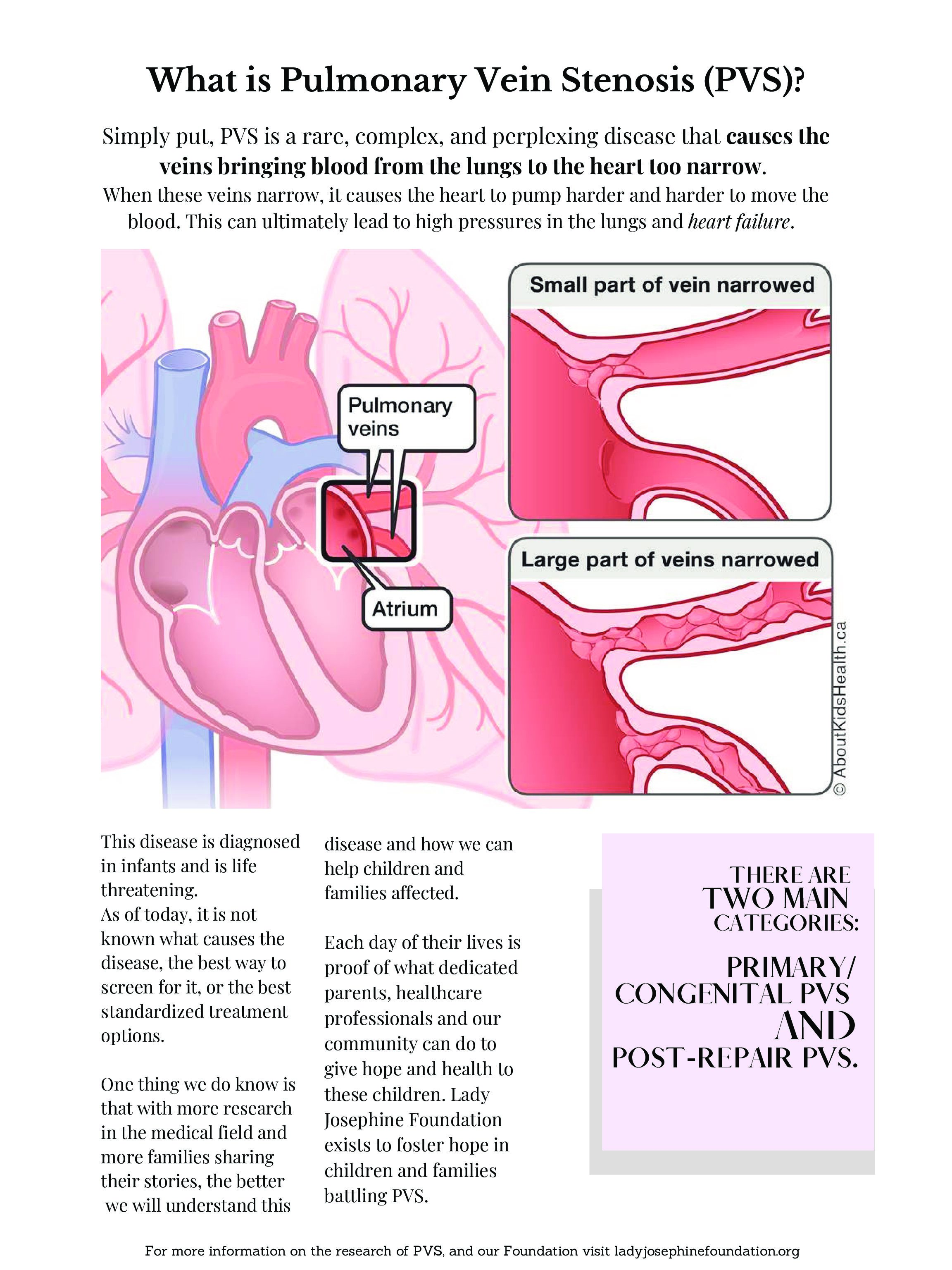
“Pediatric Pulmonary Vein Stenosis (PVS) is a devastating rare disease. Classified as a congenital heart disease, it affects the entire cardiovascular system and is generally most aggressive in early life. The road taken by parents is often filled with complications, worry, fear, trauma, endless decisions, fatigue, frustration, the search for answers. And yet, hope. Joy. An appreciation for the little things. Deep gratitude. Faith. Resilience. Strength beyond imagination.
In this podcast, we’ll dive into family stories and topics relevant to PVS life.”
Against the Odds: Improving Survival for Children with Pulmonary Vein Stenosis
Episode Summary
Pulmonary vein stenosis (PVS) is a rare and challenging condition which narrows blood vessels around the lungs and typically impacts newborn children. Historically the rates of survival have been low. But Dr. Christopher Petit has dedicated his career to developing innovative techniques and treatment options for PVS that are changing the trajectory of his patients’ lives.
Episode Notes
Dr. Christopher Petit is the co-director of the Children’s Heart Center at NewYork-Presbyterian Morgan Stanley Children’s Hospital and division chief of pediatric cardiology at Columbia University, and… a stubborn optimist. That optimism has made all the difference in his years of treating a rare heart disease in newborns: Pulmonary Vein Stenosis (PVS). Dr. Petit’s determination to better understand and treat PVS has led him and his team to make important breakthroughs in the field, including the use of sirolimus– an immune-targeted medication usually used for cancer treatments– as a systemic oral therapy to treat PVS, as well as the development of a hybrid surgical method for treating patients with severe disease, performed in collaboration with Dr. Emile Bacha. The results: drastically improved survival rates for his patients.
PVS White Papers
Systemic Sirolimus Therapy Is Associated With Reduced Intervention Frequency in Pulmonary Vein Stenosis, 2024
Kalustian, A, Hagan, J, Brlecic, P. et al. Systemic Sirolimus Therapy Is Associated With Reduced Intervention Frequency in Pulmonary Vein Stenosis. JACC Adv. 2024 Dec, 3 (12_Part_1) .
https://doi.org/10.1016/j.jacadv.2024.101401
In summary:
This article explores the use of systemic sirolimus therapy (SST) for treating pediatric pulmonary vein stenosis (PVS), a rare heart condition that causes narrowing of the veins that carry blood from the lungs to the heart. The main goal of the study was to see if SST could reduce the number of medical interventions required for PVS.
The study is different from previous research because it looks at a larger group of patients over a longer period (from 2015 to 2022). It also uses advanced statistical methods to compare the number of interventions needed before and after SST treatment. This helps provide a clearer picture of how SST might affect PVS patients in the real world.
The key finding of the study is that patients who received SST had fewer interventions compared to when they were not on the therapy. The median number of interventions per patient dropped from 5 per year before starting SST to 1.7 per year while on SST. This suggests that SST could help reduce the need for repeated surgeries or catheter procedures.
Additionally, the study found that survival rates for patients on SST were better than what’s reported in other studies, where survival is often very low after PVS interventions. However, the study didn’t include a control group, making it hard to say for sure that SST caused these better outcomes.
One concern raised by the study is the potential for infections due to the immunosuppressive nature of SST. Some patients died after infections, though it’s unclear if SST contributed to these deaths.
Overall, the study shows that SST could be an effective treatment for PVS, but it also highlights the need for further research. Future studies should have control groups, longer follow-up times, and a clearer understanding of the best duration for SST treatment.
*Neonatal Risk Factors for Pulmonary Vein Stenosis in Infants Born Preterm with Severe Bronchopulmonary Dysplasia, 2024
McArthur E, Murthy K, Zaniletti I, Sharma M, Lagatta J, Ball M, Porta N, Grover T, Levy P, Padula M, Hamrick S, Vyas-Read S; CHNC Severe BPD and Cardiology Focus Groups. Neonatal Risk Factors for Pulmonary Vein Stenosis in Infants Born Preterm with Severe Bronchopulmonary Dysplasia. J Pediatr. 2024 Dec;275:114252.Epub 2024 Aug 22. PMID: 39181320.
https://doi.org/10.1016/j.jpeds.2024.114252
Preview:
Management of Pediatric Pulmonary Vein Stenosis, 2022
Callahan, R., Morray, B. H., Hirsch, R., & Petit, C. J. (2022). Management of Pediatric Pulmonary Vein Stenosis. Journal of the Society for Cardiovascular Angiography & Interventions, 100391
https://doi.org/10.1016/j.jscai.2022.100391
In summary:
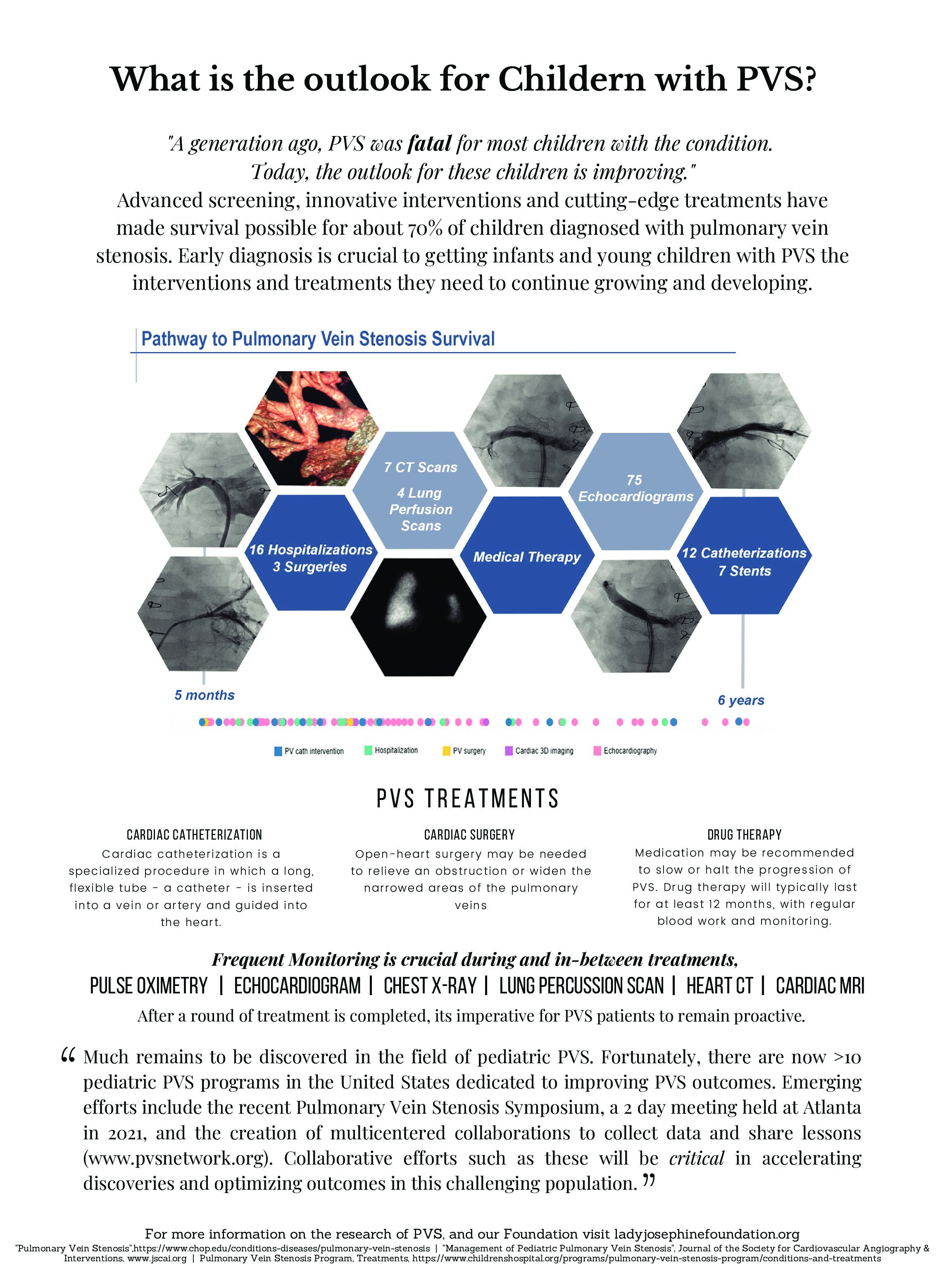
The article focuses on pediatric intraluminal pulmonary vein stenosis (PVS), a condition where the pulmonary veins in children become narrowed due to thickening of the vein walls. This causes high pressure in the lungs, leading to breathing problems and poor growth. While this disease was once considered fatal, survival rates have improved, and it is now considered a chronic illness that can be managed with proper treatment.
The article will explain the details of PVS, including how it develops, its symptoms, and how it affects children’s health. It discusses the various causes and risk factors, such as premature birth or heart defects, and how these factors can lead to the disease. It also highlights the importance of early diagnosis, surveillance, and a multi-specialty approach to treatment, as managing PVS requires the collaboration of various healthcare professionals.
The article will also cover the different types of PVS, the methods used to diagnose it (such as imaging techniques), and the treatments available, which may include surgeries or interventions to manage the narrowed veins. Finally, it will discuss the challenges of treating the condition, including the role of other health problems or comorbidities, and how these can impact the treatment outcomes.
In summary, reading the full article will give you a deeper understanding of how PVS is diagnosed, treated, and managed, and the ongoing research and strategies being developed to improve the quality of life and survival rates for children with this condition.
*Primary pulmonary vein stenosis during infancy: state of the art review, 2021
Frank, D. B., Levy, P. T., Stiver, C. A., Boe, B. A., Baird, C. W., Callahan, R. M., Smith, C. v, Vanderlaan, R. D., & Backes, C. H. (2021). Primary pulmonary vein stenosis during infancy: state of the art review. Journal of Perinatology : Official Journal of the California Perinatal Association, 41(7), 1528–1539.
https://doi.org/10.1038/s41372-021-01008-7
Preview:
Overview
Primary pulmonary vein stenosis (PPVS) is an emerging problem among infants. In contrast to acquired disease, PPVS is the development of stenosis in the absence of preceding intervention. While optimal care approaches remain poorly characterized, over the past decade, understanding of potential pathophysiological mechanisms and development of novel therapeutic strategies are increasing. A multidisciplinary team of health care providers was assembled to review the available evidence and provide a common framework for the diagnosis, management, and treatment of PPVS during infancy. To address knowledge gaps, institutional and multi-institutional approaches must be employed to generate knowledge specific to ex-premature infants with PPVS. Within individual institutions, creation of a team comprised of dedicated health care providers from diverse backgrounds is critical to accelerate clinical learning and provide care for infants with PPVS. Multi-institutional collaborations, such as the PVS Network, provide the infrastructure and statistical power to advance knowledge for this rare disease.
Pulmonary vein stenosis: Anatomic considerations, surgical management, and outcomes, 2021
Feins, E. N., Ireland, C., Gauvreau, K., Chávez, M., Callahan, R., Jenkins, K. J., & Baird, C. W. (2021). Pulmonary vein stenosis: Anatomic considerations, surgical management, and outcomes. The Journal of Thoracic and Cardiovascular Surgery.
https://doi.org/10.1016/j.jtcvs.2021.10.022
In summary:
This article discusses a study on how doctors have been treating a rare and serious condition called pulmonary vein stenosis (PVS) in children. PVS occurs when the veins that carry blood from the lungs to the heart become narrowed, which can cause breathing problems and other health issues. The study looked at the outcomes of 174 children who underwent surgery to fix this problem at a hospital over 13 years, from 2007 to 2019.
The main goal of the study was to see how different surgical techniques for fixing PVS have worked over time. Doctors have tried several methods to treat PVS, including older methods like “sutureless repair” and newer ones like a “focused anatomically-based repair.” The new approach aims to make the veins straighter and shorter, which helps reduce blood flow problems. This study showed that children who received the newer “focused repair” had better survival rates.
Some key points:
-
174 children had surgery for PVS.
-
There were different types of PVS: some kids had the condition from birth (primary), while others developed it after previous heart surgeries.
-
The children who had the anatomically focused repair surgery had better chances of survival compared to those who had older types of surgery.
-
The study also found that chemotherapy was used in some cases to help prevent the condition from coming back.
-
Overall, despite improvements in surgery, PVS is still a challenging disease to treat, and doctors need to keep improving care for kids with this condition.
What You’ll Find in the Full Article:
-
Different Treatment Methods: It goes into detail about the different surgeries used to treat PVS, such as the “sutureless” method and the newer “anatomically focused” repair. It also discusses why doctors are trying to make the veins straighter and shorter to improve blood flow.
-
Study Results: You’ll read about the outcomes of the surgeries over the years, including survival rates and the need for follow-up surgeries or treatments. It also explains how the new approach showed better survival rates in children.
-
Post-surgery Care: The article describes the care patients receive after surgery, such as follow-up tests to make sure the veins stay open, and how doctors sometimes use chemotherapy to help prevent the condition from coming back.
-
Future of Treatment: The article suggests that while the new surgery has improved outcomes, PVS is still hard to treat, and more research is needed to find the best long-term solutions.
If you read the full article, you’ll gain a deeper understanding of how doctors are working to treat a challenging heart condition in kids and how surgical techniques are evolving to improve patient outcomes.
Outcomes in Establishing Individual Vessel Patency for Pediatric Pulmonary Vein Stenosis, 2021
Callahan, R., Gauvreau, K., Marshall, A. C., Sena, L. M., Baird, C. W., Ireland, C. M., McEnaney, K., Bjornlund, E. C., Mendonca, J. T., & Jenkins, K. J. (2021). Outcomes in Establishing Individual Vessel Patency for Pediatric Pulmonary Vein Stenosis. Children (Basel, Switzerland), 8(3).
https://doi.org/10.3390/children8030210
In summary:
This study looks at how the characteristics of pulmonary veins (the blood vessels that carry blood from the lungs to the heart) and the patients themselves can predict the outcome of a disease called pulmonary vein stenosis (PVS) in children. PVS is a condition where these veins become narrow, causing issues like high blood pressure in the lungs, heart failure, and even death.
The researchers studied 48 children who were part of a clinical trial where they received treatment with two drugs (imatinib mesylate and possibly bevacizumab) after surgery to open up the narrowed veins. They focused on how the disease in each individual vein progressed over time, looking for patterns to help predict which veins were likely to do better or worse. They studied 182 individual pulmonary veins and tracked their condition over 72 weeks.
Here’s a simple breakdown of what they found:
Age and Disease Progression: Older patients (over 1 year old) tended to have better outcomes. Younger children, especially those diagnosed before 6 months, were at higher risk of the disease getting worse.
Vein Location Matters: The location of the pulmonary vein was important. Veins that were located in the left upper part of the chest tended to do worse, while veins in the right lower part did better overall.
Severity at Diagnosis: Veins that had no disease at the start were more likely to stay healthy, whereas veins with more advanced disease (especially those with problems at the far end of the vein) were more likely to require more interventions (like surgery).
Drug Treatment: How much of the prescribed drug treatment a patient received also mattered. Patients who received a higher percentage of the intended doses had fewer problems and needed fewer surgeries.
The study helps us understand that treating PVS is not just about the patient’s overall condition, but also about looking at each vein individually. Factors like age, where the vein is located, how severe the disease is at the start, and how much of the treatment the patient gets all play a big role in whether the vein improves or gets worse.
In the future, doctors may use this information to tailor treatments more specifically to each vein and patient, which could improve outcomes for kids with PVS.
Aspiration Is Associated with Poor Treatment Response in Pediatric Pulmonary Vein Stenosis, 2021
Niccum, M., Callahan, R., Gauvreau, K., & Jenkins, K. J. (2021). Aspiration Is Associated with Poor Treatment Response in Pediatric Pulmonary Vein Stenosis. Children (Basel, Switzerland), 8(9).
https://doi.org/10.3390/children8090783
In summary:
This article discusses a rare and serious condition called pulmonary vein stenosis (PVS), where the veins that carry blood from the lungs to the heart become narrowed. It can lead to severe health problems like difficulty breathing, heart failure, and even death. The article explores a specific factor, aspiration (when food or liquids accidentally enter the airway instead of going to the stomach), and how it might affect the success of treatments for PVS, particularly in children.
The study looked at 84 children with 2-ventricle heart physiology (meaning their hearts have two pumping chambers). These children received a combination of two drugs, imatinib mesylate and bevacizumab, as part of a treatment plan to manage their PVS. The study found that children who had aspiration problems were much more likely to have poor treatment outcomes, such as needing frequent re-interventions or even death. Specifically, kids with aspiration were 5 times more likely to have poor responses to treatment than those without aspiration. The study also found that male children had worse treatment outcomes compared to females.
The study suggests that aspiration may be a modifiable risk factor for poor treatment response, meaning that treating aspiration more aggressively could potentially improve the outcomes for these kids. The researchers think that aspiration might lead to lung damage, which could worsen the PVS condition.
Key Findings:
-
Aspiration (breathing in food or liquids) significantly increases the risk of poor treatment outcomes for kids with PVS.
-
Male sex was also linked to worse responses to treatment.
-
Addressing aspiration early and aggressively might improve treatment outcomes for these patients.
In conclusion, the study highlights that aspiration and male sex are important factors that should be considered when treating children with PVS. More research is needed to fully understand how aspiration affects treatment and how to best manage it to improve patient outcomes.
Systemic Sirolimus Therapy for Infants and Children With Pulmonary Vein Stenosis, 2021
Patel, J. D., Briones, M., Mandhani, M., Jones, S., Suthar, D., Gray, R., Pettus, J., McCracken, C., Thomas, A., & Petit, C. J. (2021). Systemic Sirolimus Therapy for Infants and Children With Pulmonary Vein Stenosis. Journal of the American College of Cardiology, 77(22), 2807–2818.
https://doi.org/10.1016/j.jacc.2021.04.013
In summary:
This study investigates the impact of systemic sirolimus therapy (SST) in infants and children with severe pulmonary vein stenosis (PVS), a rare and challenging condition, particularly in cases involving multivessel disease. The goal was to determine if sirolimus, an mTOR inhibitor, could improve survival and slow the progression of PVS when used as a primary medical therapy, in combination with anatomic interventions like balloon angioplasty or stent implantation.
Key Points of the Study:
-
Study Design: This was a single-center, retrospective study conducted between 2015 and 2020, focusing on infants and young children diagnosed with moderate-to-severe PVS. Out of 67 patients treated, 15 were given sirolimus, and the remaining 52 patients formed the control group. The primary endpoint was survival, and the secondary endpoint was adverse events (AEs) related to SST.
-
Results:
-
Survival Advantage: The SST group showed 100% survival compared to only 45% survival in the control group, with statistical significance (log-rank p = 0.004).
-
Sensitivity Analysis: Adjusting for survival bias by accounting for the median time from diagnosis to SST initiation confirmed that the survival advantage persisted (log-rank p = 0.027).
-
Adverse Events (AEs): Only two mild AEs were observed, both of which were treatable and did not require discontinuation of therapy.
-
Interventions: Patients in the SST group required more frequent transcatheter interventions, with a median of 3.7 catheterizations per person-year, highlighting the ongoing need for anatomic therapies even with SST.
-
-
Mechanism of Action of Sirolimus: Sirolimus targets the mTOR pathway, known to be involved in vascular cell proliferation, which is a key mechanism in the pathophysiology of PVS. The therapy is expected to slow or halt the progression of both native pulmonary vein stenosis and in-stent stenosis, which is particularly relevant for patients requiring stents.
-
Safety Profile: Sirolimus was well tolerated, with only two mild medically significant AEs observed during the study period. This safety profile aligns with previous research in pediatric transplant populations.
-
Study Limitations: This study is limited by its retrospective nature, the small sample size, and the fact that the SST group received more frequent interventions, which complicates isolating the effect of sirolimus alone. The lack of randomization also means there may be confounding factors affecting the outcomes.
-
Conclusions: The study demonstrates that systemic sirolimus therapy, when used as part of a comprehensive treatment regimen for severe PVS, significantly improves survival outcomes. Despite the need for frequent anatomic interventions, the results suggest that sirolimus plays a crucial role in altering the underlying pathophysiology of PVS, offering hope for a durable therapeutic approach. However, further studies, including randomized controlled trials, are needed to confirm these findings and better understand the optimal timing and duration of therapy.
Key Takeaways:
-
Improved Survival with Sirolimus: A survival benefit was noted in the SST cohort, even though these patients underwent more frequent interventions.
-
Safety and Tolerability: Sirolimus was generally well tolerated, with only mild AEs.
-
Need for Further Research: Given the retrospective nature of this study, future randomized controlled trials are necessary to more definitively establish the efficacy and safety of sirolimus in PVS management.
This study contributes valuable evidence supporting the use of sirolimus as a potential primary medical therapy for children with severe PVS, especially in the context of high-risk, multivessel disease.
*Systemic Sirolimus to Prevent In-Stent Stenosis in Pediatric Pulmonary Vein Stenosis, 2020
Callahan, R., Esch, J. J., Wang, G., Ireland, C. M., Gauvreau, K., & Jenkins, K. J. (2020). Systemic Sirolimus to Prevent In-Stent Stenosis in Pediatric Pulmonary Vein Stenosis. Pediatric Cardiology, 41(2), 282–289.
https://doi.org/10.1007/s00246-019-02253-6
Preview:
Overview
Evaluate the efficacy of systemic sirolimus (rapamycin) in preventing in-stent stenosis (ISS) in pediatric intraluminal pulmonary vein stenosis (PVS). Report the adverse events related to sirolimus therapy. There is a high incidence of ISS following stent implantation in PVS. The use of sirolimus in preventing ISS has not been reported. Retrospective review of all patients who received sirolimus (8 week course) for treatment of ISS for PVS between January 2013 and June 2018. Forty stents (37 bare metal, 3 drug-eluting) in 20 patients were treated with sirolimus; 20 at the time of implantation (primary prevention [1P]) and 20 following documented ISS requiring transcatheter reintervention (secondary prevention [2P]). Treated patients were young (median 2 y/o [0.7–5.7]) and most had PVS associated with congenital heart disease (75%, 15/20; 4/15 with TAPVC). In the 1P group, 85% (17/20) of stents were without significant (< 50%) ISS at median of 102 days (range 56–527); the growth rate of ISS in this group was 7.5 ± 7.1%/month. In the 2P group, most stents had a slower growth rate of ISS after sirolimus therapy compared to pre-treatment (median 3.7 [− 0.2 to 13.1] vs. 10.4 [1.3 to 19.5] %/month; p < 0.001). One patient developed pneumonia on drug while concurrently taking another immunosuppressive agent. No other serious adverse events were related to sirolimus therapy. Systemic sirolimus slows the growth rate of ISS following stent implantation in PVS compared to pre-treatment rates and was administered safely in a small number of pediatric patients with complex heart disease.
*Comparison of drug eluting versus bare metal stents for pulmonary vein stenosis in childhood, 2019
Khan, A., Qureshi, A. M., & Justino, H. (2019). Comparison of drug eluting versus bare metal stents for pulmonary vein stenosis in childhood. Catheterization and Cardiovascular Interventions, 94(2), 233–242.
https://doi.org/10.1002/ccd.28328
Preview:
Objective
Comparison of outcomes using bare metal (BMS) and drug-eluting (DES) stents in pulmonary vein stenosis (PVS).
Background
PVS is a serious condition with frequent restenosis after surgical and percutaneous interventions. After experiencing encouraging results with DES, we sought to compare outcomes of BMS and DES in native and post-surgical PVS.
Methods and Results
A retrospective review of all patients who underwent stent implantation between 08/93 and 11/17 for PVS at Texas Children’s Hospital was performed. BMS were used to treat 58 lesions in 37 patients and 105 DES used to treat 105 lesions in 41 patients. Mean age at first stent implant was 2.9 ± 3.5 years in BMS and 16.2 ± 18.8 months in DES group. Of those with follow-up catheterization, mean lumen loss rate from stent implant to first follow-up catheterization was 0.85 ± 1.47 mm/month over 6.4 ± 6.4 months in the BMS group (n = 44 lesions) compared to 0.16 ± 0.31 mm/month over 6.8 ± 7.4 months in the DES group (n = 86 lesions), p < .01. Follow-up for the BMS group was 14 months (6 days–22.3 years), with 13 mortalities, eight lesions were re-stented and six complete occlusions were noted. Follow-up for DES group (including four cross-overs) was 17.5 months (3 days-9 years), with 10 mortalities, seven lesions were re-stented, 11 had complete occlusion, 20 new adjacent lesions in the same vessel underwent stenting and 12 stents were intentionally fractured.
Conclusion
DES have significantly lowered lumen loss rate when compared to BMS at medium term follow-up and can be fractured to enable larger diameters. Availability of larger diameter DES would be ideal.
*Pulmonary Vein Stenosis: Outcomes in Children With Congenital Heart Disease and Prematurity, 2019
DiLorenzo, M. P., Santo, A., Rome, J. J., Zhang, H., Faerber, J. A., Mercer-Rosa, L., & Hopper, R. K. (2019). Pulmonary Vein Stenosis: Outcomes in Children With Congenital Heart Disease and Prematurity. Seminars in Thoracic and Cardiovascular Surgery, 31(2), 266–273.
https://doi.org/10.1053/j.semtcvs.2018.09.027
Preview:
*Adjunct Targeted Biologic Inhibition Agents to Treat Aggressive Multivessel Intraluminal Pediatric Pulmonary Vein Stenosis, 2018
Callahan, R., Kieran, M. W., Baird, C. W., Colan, S. D., Gauvreau, K., Ireland, C. M., Marshall, A. C., Sena, L. M., Vargas, S. O., & Jenkins, K. J. (2018). Adjunct Targeted Biologic Inhibition Agents to Treat Aggressive Multivessel Intraluminal Pediatric Pulmonary Vein Stenosis. The Journal of Pediatrics, 198, 29-35.e5.
https://doi.org/10.1016/j.jpeds.2018.01.029
Preview:
Reintervention Is Associated With Improved Survival in Pediatric Patients With Pulmonary Vein Stenosis, 2017
Cory, M. J., Ooi, Y. K., Kelleman, M. S., Vincent, R. N., Kim, D. W., & Petit, C. J. (2017). Reintervention Is Associated With Improved Survival in Pediatric Patients With Pulmonary Vein Stenosis. JACC: Cardiovascular Interventions, 10(17), 1788–1798.
https://doi.org/10.1016/j.jcin.2017.05.052
In summary:
The article discusses a study on pulmonary vein stenosis (PVS), a rare condition in children where the veins carrying blood from the lungs to the heart become narrowed, potentially causing heart failure and breathing problems. The study aimed to evaluate the survival rates of children who underwent catheter-based treatments for PVS, including balloon angioplasty, bare-metal stents (BMS), and drug-eluting stents (DES). The study found that, of the 30 children treated, 47% died within a few months, mostly due to complications from PVS. However, children who had repeat treatments (such as additional catheter procedures or surgery) had a significantly better chance of surviving, with a 1-year survival rate of 84% for those who received further treatment, compared to just 25% for those who didn’t.
While many of the treated veins were lost over time, the study found that DES, which slowly releases medicine to help prevent vein re-blockage, was more effective in keeping veins open longer compared to other treatments. However, DES did not improve the children’s overall survival, meaning that while it helps the veins stay clear, it might not directly affect how long the child lives. The study also emphasized that frequent follow-up and early treatment of recurring stenosis were critical for improving survival rates. Despite the positive outcomes with repeated treatments, the study also noted limitations, as it was based on past cases, and doctors made different treatment choices. The researchers suggest that future studies should explore newer treatment options, like drug-eluting balloons, and involve multiple hospitals to better understand the most effective ways to treat PVS in children.
Overall, the study concluded that while DES may help with vein survival, regular check-ups and reinterventions are essential for improving the chances of long-term survival in children with PVS.
*Paucicellular Fibrointimal Proliferation Characterizes Pediatric Pulmonary Vein Stenosis, 2017
Kovach, A. E., Magcalas, P. M., Ireland, C., McEnany, K., Oliveira, A. M., Kieran, M. W., Baird, C. W., Jenkins, K., & Vargas, S. O. (2017). Paucicellular Fibrointimal Proliferation Characterizes Pediatric Pulmonary Vein Stenosis: Clinicopathologic Analysis of 213 Samples From 97 Patients. The American Journal of Surgical Pathology, 41(9), 1198–1204.
https://doi.org/10.1097/PAS.0000000000000892
Preview:
Overview
Pulmonary vein stenosis (PVS) is a luminal narrowing of extrapulmonary pulmonary veins. In pediatric patients, it arises following repair of congenital heart disease, particularly anomalous pulmonary venous return; in lung disease, especially prematurity; and rarely in isolation. The etiology is unknown and the course often fatal without lung transplantation. We hypothesized that systematic clinicopathologic review of pediatric PVS could provide further pathogenic insight. We included patients who underwent first resection of pulmonary venous tissue for symptomatic PVS at our pediatric referral center from 1995 to 2014. Clinical records and hematoxylin and eosin slides were reviewed. Subsets were immunostained for smooth muscle actin, Ki-67, β-catenin, estrogen receptor, and other markers and analyzed for USP6 gene rearrangement. A total of 97 patients (57% male; median age: 6 mo) were identified. Overall, 59 (61%) had prior congenital heart disease repair, 35 involving pulmonary vein manipulation. Samples included 213 separate anatomic sites (median: 2/patient). Histologically, all showed sparsely cellular intimal expansion composed of haphazardly arranged fibroblasts with slender nuclei in myxoid matrix. This tissue merged with underlying collagen. Most samples had a variably continuous sheath of cardiomyocytes. Ancillary tests supported a reactive fibroblastic proliferation; in particular, fibroblasts showed cytoplasmic β-catenin localization, no estrogen receptor expression, and no USP6 rearrangement. At last follow-up (mean: 2.3 y), 46% of patients had died of disease. Pediatric PVS uniformly consists of a paucicellular fibrointimal proliferation, irrespective of clinical scenario. It may be best conceived of as a form of reactive hyperplasia. As with other forms of vascular remodeling, trauma (iatrogenic or occult) is likely an inciting factor. A comprehensive understanding of the surgical pathology of PVS may further inform therapeutic strategies in this lethal disease.
Primary Pulmonary Vein Stenosis: Outcomes, Risk Factors, and Severity Score in a Multicentric Study, 2017
Kalfa, D., Belli, E., Bacha, E., Lambert, V., di Carlo, D., Kostolny, M., Salminen, J., Nosal, M., Poncelet, A., Horer, J., Berggren, H., Yemets, I., Hazekamp, M., Maruszewski, B., Sarris, G., Pozzi, M., Ebels, T., Lacour-Gayet, F., & European Congenital Heart Surgeons Association. (2017). Primary Pulmonary Vein Stenosis: Outcomes, Risk Factors, and Severity Score in a Multicentric Study. The Annals of Thoracic Surgery, 104(1), 182–189.
https://doi.org/10.1016/j.athoracsur.2017.03.022
In summary:
The article discusses a study on primary pulmonary vein stenosis (PPVS), a rare heart condition in which the veins that carry blood from the lungs to the heart become narrowed, making it difficult for the heart to function properly. The study’s goal was to understand the outcomes and factors that influence survival and recovery after surgery for PPVS. It reviewed 30 patients who underwent surgery at various hospitals between 2000 and 2012. The study specifically looked at how different surgical techniques, including the sutureless technique, impacted recovery.
The results showed that PPVS remains a challenging condition to treat, with a high risk of complications. After surgery, 30% of the patients died, 40% needed another surgery, and 50% experienced restenosis (the veins narrowed again). On average, patients had about a 70% chance of surviving without needing more surgery or experiencing restenosis over 8 years. However, the study also highlighted that children who had more severe disease or developed severe pulmonary hypertension (high blood pressure in the lungs) after surgery faced worse outcomes.
The study also developed a severity score to predict how severe the condition was in each patient. The score considered factors like how much the veins were narrowed and whether the problem affected one or both veins. This score turned out to be a useful tool for predicting which patients might do poorly after surgery. Surprisingly, the type of surgery (sutureless or not) didn’t significantly affect the outcomes, meaning the severity of the disease before surgery was a much bigger factor.
In conclusion, even with advances like the sutureless surgery technique, the prognosis for PPVS remains poor. The study suggests that factors such as the severity of the disease before surgery and the presence of pulmonary hypertension afterward are strong predictors of whether a patient will recover well. More research is needed to better understand how to treat this condition effectively.
Pulmonary vein stenosis: Severity and location predict survival after surgical repair, 2016
lo Rito, M., Gazzaz, T., Wilder, T. J., Vanderlaan, R. D., van Arsdell, G. S., Honjo, O., Yoo, S.-J., & Caldarone, C. A. (2016). Pulmonary vein stenosis: Severity and location predict survival after surgical repair. The Journal of Thoracic and Cardiovascular Surgery, 151(3), 657-66.e1-2.
https://doi.org/10.1016/j.jtcvs.2015.08.121
In summary:
The article focuses on understanding the survival rates of patients who undergo surgery to repair pulmonary vein stenosis (a condition where the blood vessels that carry blood from the lungs to the heart become narrowed or blocked). The study looks at how specific measurements of the pulmonary veins before surgery (using CT and MRI scans) might predict how well patients will do after the surgery.
Here’s a simplified breakdown of what’s going on:
Background: Pulmonary vein stenosis is a serious condition that can happen either after heart surgery or as a primary birth defect. Even though surgery is often done to repair this condition, the survival rates for these patients are not very high, and surgical outcomes haven’t improved much over time.
Study Purpose: The study aims to figure out if certain features of the pulmonary veins, as seen on CT and MRI scans, can predict whether a patient will survive after surgery to fix the stenosis. These features include the size of the veins at different points (upstream, downstream, and at the narrowest part), and how these sizes might be related to survival.
Key Findings:
Smaller veins = worse survival: Patients who had smaller veins before surgery, especially in the area before the veins join the heart (upstream veins), had a higher risk of dying within the first year after surgery.
More veins affected = worse survival: Having more pulmonary veins affected by stenosis also made survival more difficult.
Flow issues: Some patients showed that blood flow was redistributed to other parts of the lungs because of the narrowing, which could indicate how well their lungs were compensating for the stenosis. This flow pattern was linked to better survival outcomes in some cases.
Conclusion: The study found that measurements of the veins’ sizes (like how wide they are) before surgery can help doctors predict which patients are at higher risk of dying after surgery. This could help with better decision-making for surgeries and potential treatments. For example, patients with smaller veins before surgery might need more intense monitoring or other treatments to improve their chances of survival.
In simpler terms: If doctors can measure the pulmonary veins before surgery and notice that they’re too small or if there are many veins affected, they might predict a poorer survival rate after the operation. This could help them decide which patients might benefit more from surgery or from trying new treatments aimed at improving vein health.
Primary pulmonary vein stenosis: the impact of sutureless repair on survival, 2011
Viola, N., Alghamdi, A. A., Perrin, D. G., Wilson, G. J., Coles, J. G., & Caldarone, C. A. (2011). Primary pulmonary vein stenosis: the impact of sutureless repair on survival. The Journal of Thoracic and Cardiovascular Surgery, 142(2), 344–350.
https://doi.org/10.1016/j.jtcvs.2010.12.004
In summary:
Study Focus:
This study focuses on primary pulmonary vein stenosis (PVS), a rare but severe condition where the veins from the lungs to the heart become narrowed or blocked. The condition can lead to high pulmonary pressure and heart failure, often resulting in early death. Over the past 20 years, medical professionals have developed a newer surgical technique called the sutureless repair to treat this condition. Unlike traditional surgery, which involves stitching the veins directly to the heart, the sutureless technique avoids contact with the vein walls, which can reduce the risk of the veins narrowing again (restenosis). What sets this study apart is its focus on whether the sutureless technique improves outcomes for patients with PVS compared to older surgical methods.
Method of the Study:
The study analyzed hospital records of 23 patients who underwent surgery for primary PVS between 1989 and 2008. The researchers reviewed the patients’ condition before surgery and followed their outcomes afterward. A key part of the analysis was the use of the PVS score, which measures the severity of the vein narrowing. Each vein in the body was given a score based on how blocked it was, and the higher the score, the worse the condition. The study aimed to see whether the new sutureless technique could improve survival rates and reduce the need for follow-up surgeries compared to older techniques, which had often failed to provide long-term relief.
Key Differences in the Approach:
A major aspect that sets this study apart is its detailed tracking of how well the sutureless technique performed in treating primary PVS. While the sutureless technique had been used before for other types of vein problems, this was one of the first studies to specifically evaluate its use for primary PVS. The researchers wanted to see if avoiding the vein wall with the sutureless technique would prevent restenosis (narrowing of the veins again after surgery), a common issue with previous surgeries. They also used a well-defined PVS score system to measure how severe the condition was before and after the surgery, which helped them understand how effective the treatments were over time.
Results:
While the sutureless technique showed some promise, the study found that mortality rates (death rates) were still very high—nearly 50% of patients died within 1 to 10 years after surgery. Interestingly, even when the sutureless technique was used, many patients still experienced restenosis, requiring additional surgeries. What made this study particularly insightful was the observation that patients with higher preoperative PVS scores (indicating more severe vein blockages) had worse outcomes, including higher rates of death and restenosis. This was a critical finding because it suggested that the severity of the disease before surgery plays a huge role in the patient’s survival and recovery, and the sutureless technique didn’t necessarily overcome this challenge.
Conclusion and New Insights:
The findings of this study suggest that while the sutureless technique might be helpful, it is not a cure-all. The study highlighted that even with this advanced technique, patients with more severe cases of PVS (those with a PVS score greater than 4) had poorer outcomes. This adds to the existing knowledge that primary PVS is a very difficult condition to treat. The use of the PVS score as a predictor of survival was another major takeaway. The study demonstrated that the PVS score can be a reliable way for doctors to predict which patients are more likely to survive after surgery, helping guide clinical decisions and set realistic expectations for treatment.
In summary, what makes this study stand out is its focus on a newer surgical technique—sutureless repair—and its use of a quantitative tool, the PVS score, to assess the severity of the condition. The research provides deeper insight into how both of these factors contribute to patient outcomes, showing that while the sutureless technique can improve surgery results for some, it still doesn’t solve the overall challenge of high mortality and restenosis, particularly in patients with more severe disease.
PVS Heros
PVS NETWORK
FOSTERING HOPE THROUGH KNOWLEDGE
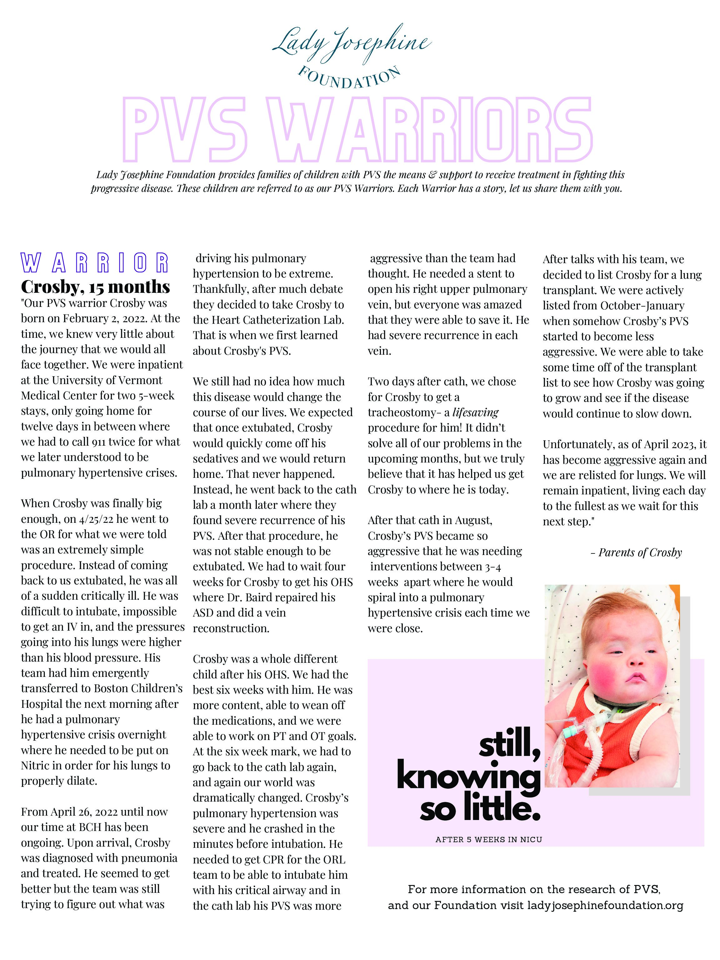
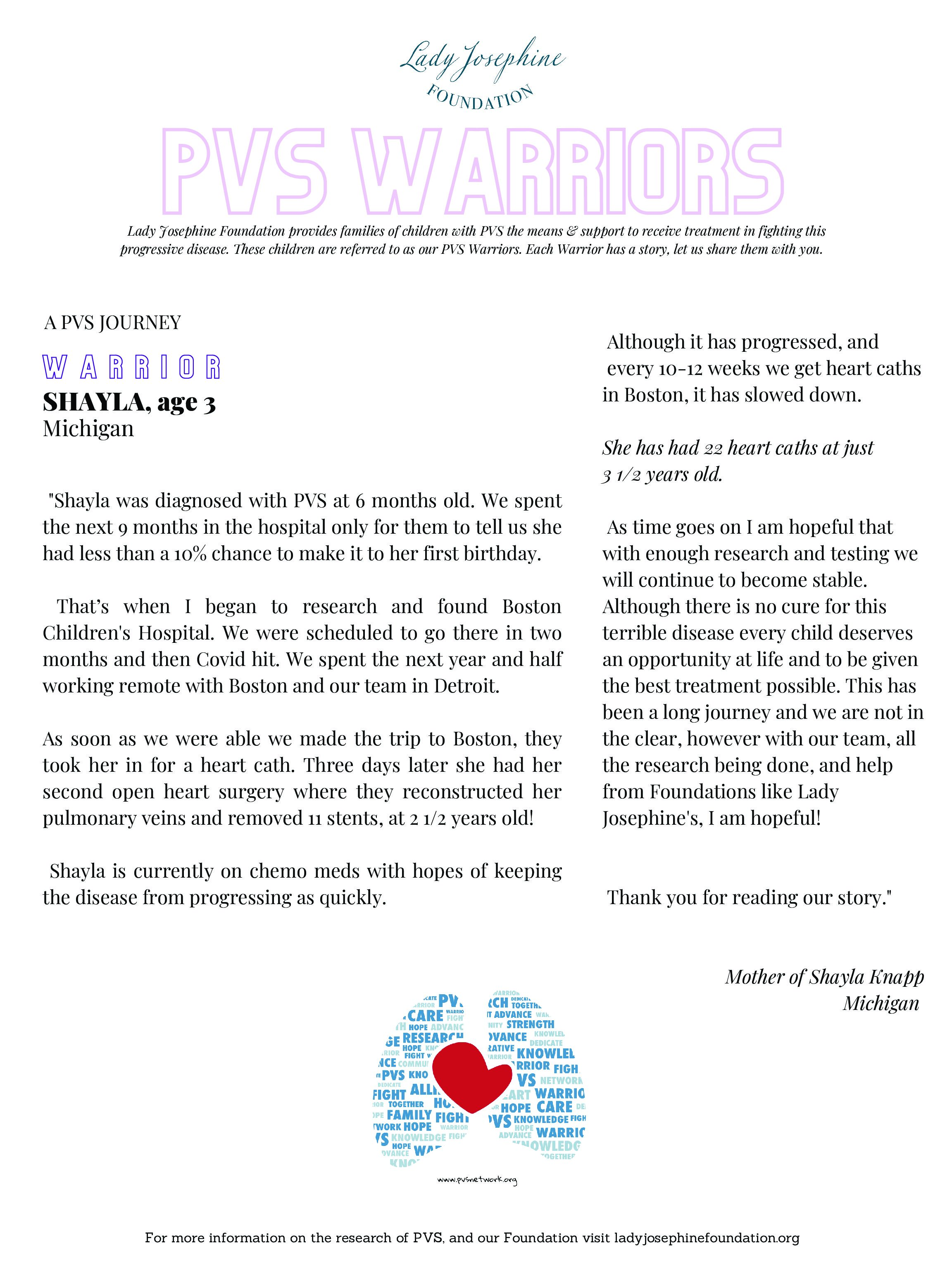
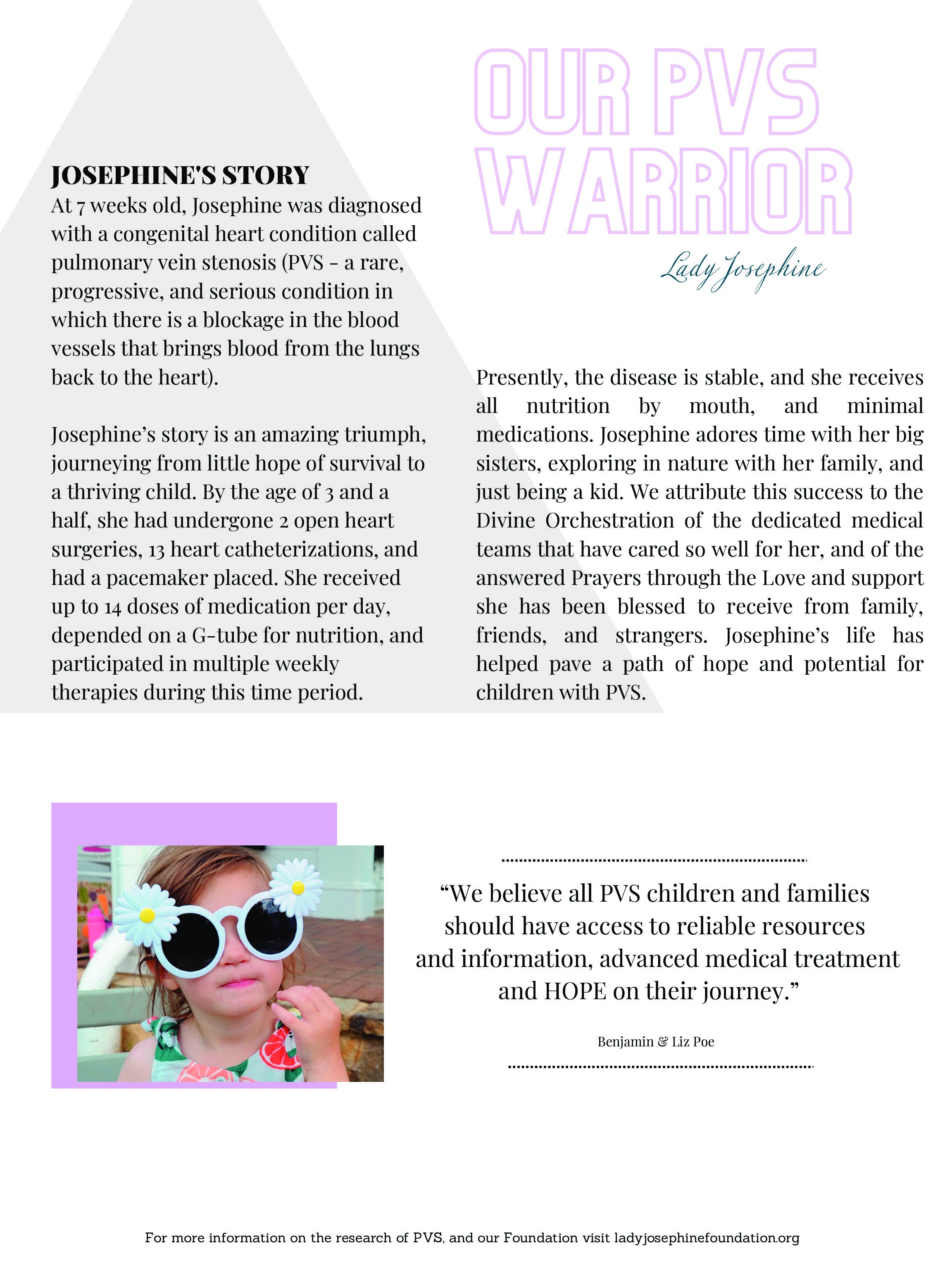
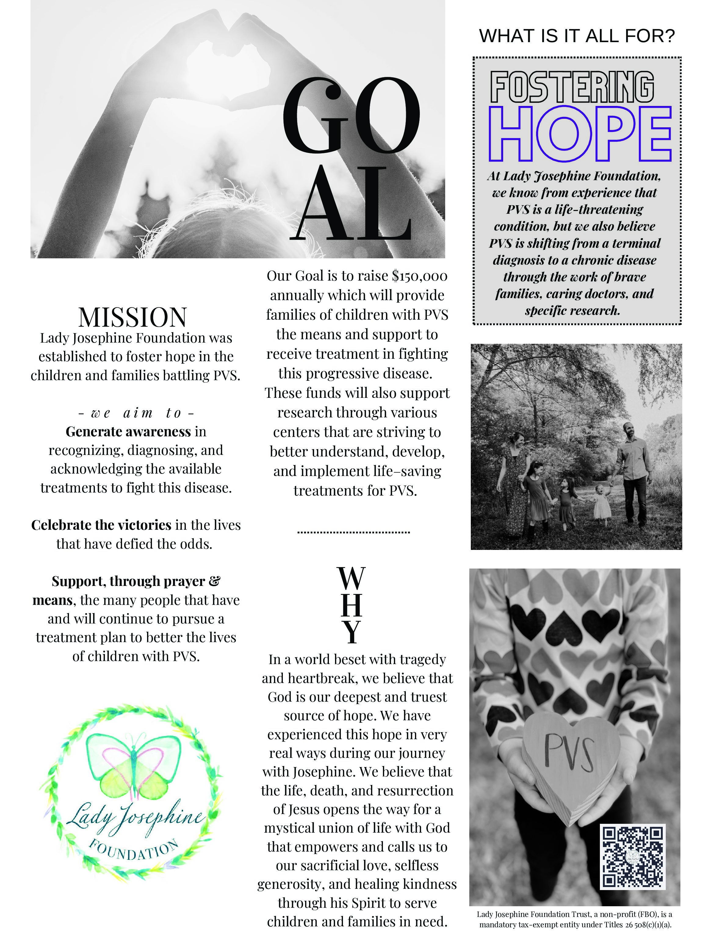
The Lady Josephine Foundation was established to acknowledge our fight for answers that seemed unattainable, to celebrate our victories in a life that has defied the odds, and to support the many people that have and will continue to pursue a treatment plan to better the lives of children with PVS. We have created the Story Boards below to help raise awareness and foster knowledge on this progressive disease. As you click through each image, you will find yourself immersed in a world where life is the goal. While better understanding PVS, you will also get to know a few of our PVS families that your contributions can directly help.
Donate Now
Make a fit to help make PVS kids better today and healthier tomorrow.